- Panchashil Plaza, 9th Floor, 55 Hughes Road, Opp. Metro Motors, Behind Dharam Palace, Mumbai - 400 007
- 9969425673

2 D Echocardiography with Colour Doppler & strain Imaging
2 D Echocardiography with Colour Doppler & strain Imaging
2D Echocardiography with Color Doppler and Strain Imaging is an advanced imaging technique used to assess the heart’s structure and function. The 2D echocardiography provides real-time, two-dimensional images of the heart, allowing doctors to visualize the chambers, valves, and walls to detect abnormalities in size, shape, or movement. The Color Doppler function adds color to these images, showing the direction and speed of blood flow through the heart. This helps identify issues like valve leakage, blockages, or abnormal blood flow patterns. Strain Imaging, on the other hand, measures the deformation or “strain” of heart muscle fibers during contraction and relaxation. It provides detailed insights into heart muscle function, enabling the detection of early signs of heart dysfunction, such as heart failure or cardiomyopathy, before they are visible on standard tests. Together, these techniques offer a comprehensive evaluation of heart health, aiding in accurate diagnosis and effective treatment planning for a range of heart conditions.
Here's a breakdown of the components and functionality of 2 D Echocardiography with Colour Doppler & strain Imaging
The functionality of 2D Echocardiography with Color Doppler and Strain Imaging combines multiple techniques to provide a comprehensive assessment of heart health:
2D Echocardiography: This feature captures two-dimensional, real-time images of the heart’s chambers, valves, and walls. It allows for the visualization of heart structures, showing their size, shape, and movement. This helps in identifying abnormalities such as valve malfunctions, chamber enlargement, or heart muscle weakness.
Color Doppler: This functionality adds color to the 2D images, visualizing blood flow within the heart. It shows the direction and speed of blood flow, helping detect conditions like valve regurgitation (backflow), stenosis (narrowing), or abnormal blood flow between chambers, aiding in the diagnosis of valve diseases and heart defects.
Strain Imaging: This measures the deformation (strain) of the heart muscle during contraction and relaxation, particularly assessing how much the heart muscle fibers stretch and contract. It provides detailed information about the heart’s mechanical function, helping to detect early signs of heart muscle dysfunction, even before visible structural damage is evident.
Together, these functionalities provide a detailed evaluation of the heart’s structure, function, and blood flow, making it an essential tool for diagnosing and monitoring heart conditions.
2 D Echocardiography with Colour Doppler & strain Imaging Procedure
The procedure for 2D Echocardiography with Color Doppler and Strain Imaging is non-invasive and typically performed in a clinical setting.
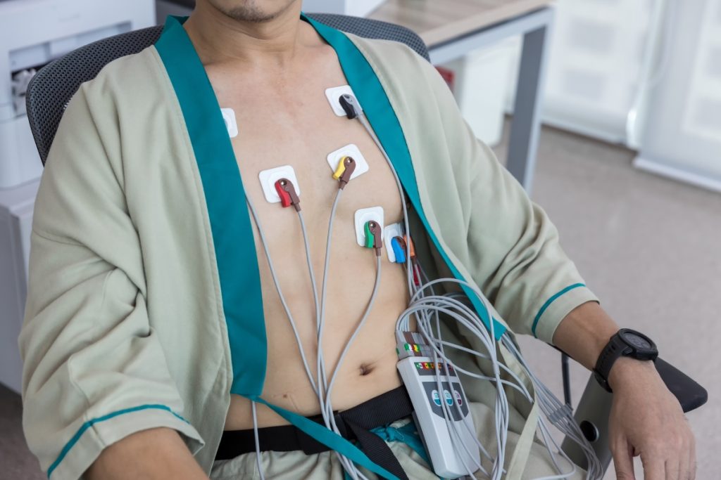
Preparation: The patient is asked to lie on an examination table, usually on their left side. A technician applies a special gel to the chest area to enhance sound wave transmission.
Transducer Placement: A transducer (ultrasound probe) is placed on the patient’s chest. The technician moves the transducer around to capture different views of the heart. The sound waves from the transducer bounce off the heart structures, creating images.
2D Echocardiography: The machine processes the sound waves to produce two-dimensional images of the heart in real-time. This allows visualization of heart chambers, valves, and walls to assess their size, shape, and motion.
Color Doppler: The Doppler function is activated, adding color to the image to visualize blood flow through the heart. It shows the speed and direction of blood flow, highlighting any abnormalities like valve leakage or blockages.
Strain Imaging: The transducer also captures data for strain imaging. Specialized software tracks the movement and deformation of heart muscle fibers to measure the strain, providing a detailed analysis of heart muscle function.
The procedure usually takes 30 to 60 minutes, is painless, and provides valuable information for diagnosing and monitoring heart conditions.

Dr. Dhiren R Shah, a highly experienced cardiologist since 1987, offers comprehensive cardiac care at his clinic. Equipped with advanced facilities
Copyright ©2024 All rights reserved | Designed by Rebecca Digital